Finding a knot on the inside of your thigh can be an unsettling experience. Whether it appeared overnight or developed gradually, any unusual mass or lump warrants a careful evaluation. In medical terms, this phenomenon refers to a localized swelling, nodule, or palpable thickening within the soft tissues, lymph nodes, or vascular structures of the medial thigh region. As medical diagnostic standards evolve through 2026, clinicians increasingly emphasize differentiating between benign superficial changes and deep-seated structural abnormalities. This comprehensive guide explores the anatomical possibilities, clinical evaluation processes, and standard care pathways associated with a knot on the inside of the thigh.
Anatomical Landscape of the Medial Thigh
The inner thigh is a complex anatomical zone packed with muscles, blood vessels, lymph nodes, and nerves. When a lump or knot forms in this area, its exact physical characteristics—such as mobility, texture, tenderness, and depth—frequently point toward specific anatomical structures.
The adductor muscle group, which includes the adductor longus, adductor brevis, adductor magnus, gracilis, and pectineus, spans the pelvis to the femur. Strains, tears, or localized fiber bunching within these muscles can create firm, knot-like areas often referred to as myofascial trigger points or muscle contraction knots. Additionally, the femoral triangle resides in the upper medial thigh, housing the femoral artery, femoral vein, and femoral nerve, alongside deep inguinal lymph nodes.
Evaluating the physical nature of the lump helps medical professionals narrow down potential causes:
- Superficial lumps located directly under the skin are frequently benign cysts or lipomas.
- Firm, fixed masses deep within the tissue layers require more urgent diagnostic imaging to rule out vascular or neoplastic concerns.
- Tender, swollen nodes localized near the groin fold typically signal an active immune response or regional infection.
Primary Clinical Causes of Inner Thigh Lumps
A knot on the inside of the thigh can stem from a wide variety of medical conditions. Understanding these possibilities helps patients prepare for meaningful discussions with their healthcare providers.
Swollen Lymph Nodes (Lymphadenopathy)
The inguinal lymph nodes sit near the crease where the abdomen meets the thigh. These nodes filter lymphatic fluid from the lower extremities, external genitalia, and lower abdominal wall. When an infection, inflammation, or injury occurs in these regions, the nearby lymph nodes can swell significantly, presenting as tender knots on the upper inside thigh. Common triggers include lower limb cellulitis, ingrown hairs, fungal infections like tinea cruris, or systemic viral illnesses.
Lipomas and Epidermoid Cysts
Lipomas are slow-growing, benign fatty tumors that sit just beneath the skin. They typically feel soft, doughy, and easily movable under gentle finger pressure. Epidermoid cysts, formed from trapped epidermal cells, present as firm, movable nodules that may occasionally discharge a cheese-like material or become inflamed.
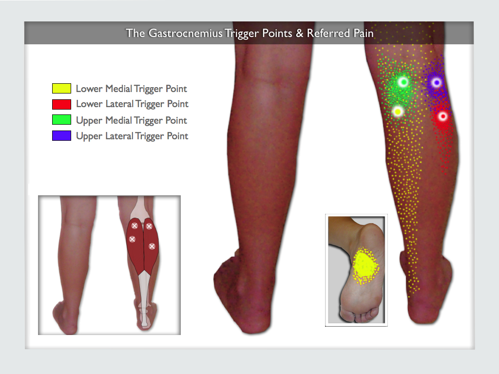
Muscle Strains and Myofascial Trigger Points
Athletes and active individuals frequently experience knots in the adductor muscles due to repetitive strain, sudden acceleration, or overstretching. These are not true structural masses, but rather hyperirritable spots within a taut band of skeletal muscle fibers that can cause localized pain and restricted range of motion.
Hernias (Femoral and Inguinal)
A hernia occurs when an internal organ or fat tissue pushes through a weak spot in the surrounding muscle or connective tissue wall. While inguinal hernias usually present in the groin, femoral hernias push down into the femoral canal, often appearing as a distinct bulge or knot just below the groin crease on the upper inner thigh. Femoral hernias carry a higher risk of strangulation and require prompt medical assessment.
Vascular Anomalies and Hematomas
Trauma to the inner thigh can rupture small blood vessels, causing blood to pool in the tissue and form a firm, painful hematoma. Less commonly, vascular conditions such as superficial thrombophlebitis (inflammation of a surface vein with a small blood clot) can present as a cord-like, tender knot running along the path of a blood vessel.
Comparative Overview of Inner Thigh Masses
To help visualize how different conditions compare in clinical presentation, the following table outlines key characteristics of common inner thigh lumps.
| Condition | Typical Texture | Mobility | Associated Pain | Primary Diagnostic Focus |
|---|---|---|---|---|
| Inguinal Lymphadenopathy | Firm, rubbery | Movable | Tender to touch | Identifying regional infection or inflammation |
| Lipoma | Soft, doughy | Highly mobile | Generally painless | Observation or simple excision if symptomatic |
| Femoral Hernia | Firm, reducible | Fixed or semi-fixed | Aching or pressure | Surgical evaluation to prevent incarceration |
| Adductor Muscle Knot | Taut, rope-like | Fixed in muscle | Achy, worsens with movement | Physical therapy and myofascial release |
| Hematoma | Firm, bruised | Fixed | Moderate to severe | Monitoring resorption or surgical drainage if large |
Diagnostic Workup and Clinical Evaluation Standards
When you present to a clinic or healthcare network with an unexplained mass on your inner thigh, providers follow a structured diagnostic framework. Modern clinical protocols prioritize non-invasive imaging alongside a thorough physical examination.
| Diagnostic Phase | Standard Procedure | Clinical Objective |
|---|---|---|
| Phase 1 | Comprehensive History & Physical Exam | Assess duration, growth rate, pain profile, and systemic symptoms like fever or unexplained weight loss. |
| Phase 2 | Diagnostic Ultrasound | Differentiate between solid masses, fluid-filled cysts, vascular structures, and hernias using high-frequency sound waves. |
| Phase 3 | Advanced Imaging (MRI/CT) | Map deep tissue involvement, evaluate complex muscle tears, or assess suspicious soft-tissue masses. |
| Phase 4 | Biopsy or Lab Work | Analyze tissue architecture via core needle biopsy or evaluate inflammatory markers through blood tests when infection is suspected. |
Navigating Healthcare Networks and Insurance Coverage
When seeking specialized care for an undiagnosed mass, understanding your insurance network and facility affiliations ensures seamless care coordination. For patients utilizing managed care systems or regional health networks:
- Ensure your chosen provider is contracted within your specific insurance tier to avoid out-of-pocket surprise billing.
- Verify whether your health plan requires an explicit primary care physician (PCP) referral before scheduling a consultation with a general surgeon, vascular specialist, or orthopedic expert.
- Confirm that diagnostic facilities—such as outpatient imaging centers—are fully accredited and recognized within your insurance provider network.
Red Flag Symptoms Requiring Immediate Medical Attention
While many inner thigh lumps are benign and resolve with conservative care or minor intervention, certain symptoms indicate a medical emergency. Seek immediate evaluation at an emergency department if you experience:
- Sudden, severe, and worsening pain accompanied by a firm, irreducible lump in the upper inner thigh or groin.
- Nausea, vomiting, or abdominal cramping alongside the thigh mass, which strongly suggests a strangulated hernia.
- Rapid enlargement of the lump, accompanied by skin discoloration, warmth, and systemic fever.
- Sudden swelling of the entire lower leg, which may indicate a deep vein thrombosis (DVT).
Frequently Asked Questions
What does it mean if I have a painful knot on the inside of my thigh?
A painful knot on the inside of the thigh most commonly indicates an inflamed lymph node, a muscle strain with a trigger point, or an infected cyst. Immediate evaluation by a healthcare professional is necessary to rule out more serious vascular or structural issues.
Could an inner thigh knot be a hernia?
Yes, a femoral or inguinal hernia can present as a noticeable bulge or knot in the upper inner thigh or groin crease. These require professional medical diagnosis because they carry a risk of tissue entrapment or strangulation.
How do doctors diagnose the cause of a thigh lump?
Doctors typically begin with a physical examination followed by a diagnostic ultrasound to visualize whether the lump is solid, fluid-filled, vascular, or muscular. Further imaging like an MRI or a tissue biopsy may be ordered if ambiguity remains.
When should I worry about a lump on my leg?
You should schedule an urgent medical evaluation if the lump is growing rapidly, feels completely fixed and hard, causes severe pain, or is accompanied by systemic signs like fever, unexplained weight loss, or spreading skin redness.
Can muscle knots form in the inner thigh?
Yes, the adductor muscles of the inner thigh frequently develop myofascial trigger points or knots due to athletic overexertion, sudden strains, or chronic postural imbalances.
Are all fatty lumps on the thigh cancerous?
No, the vast majority of superficial fatty lumps are benign lipomas, which are non-cancerous growths of fat cells. However, any new or changing mass should be formally evaluated to confirm its benign nature.
Professional Care and Consultation
Addressing an unexplained knot on the inside of your thigh begins with a professional clinical evaluation. Delaying diagnosis can complicate treatment pathways, particularly if the mass is a hernia or an evolving soft-tissue condition. Schedule an appointment with your primary care provider or an appropriate specialist today to receive an accurate assessment, rule out serious pathology, and establish a personalized, effective treatment plan tailored to your health needs.