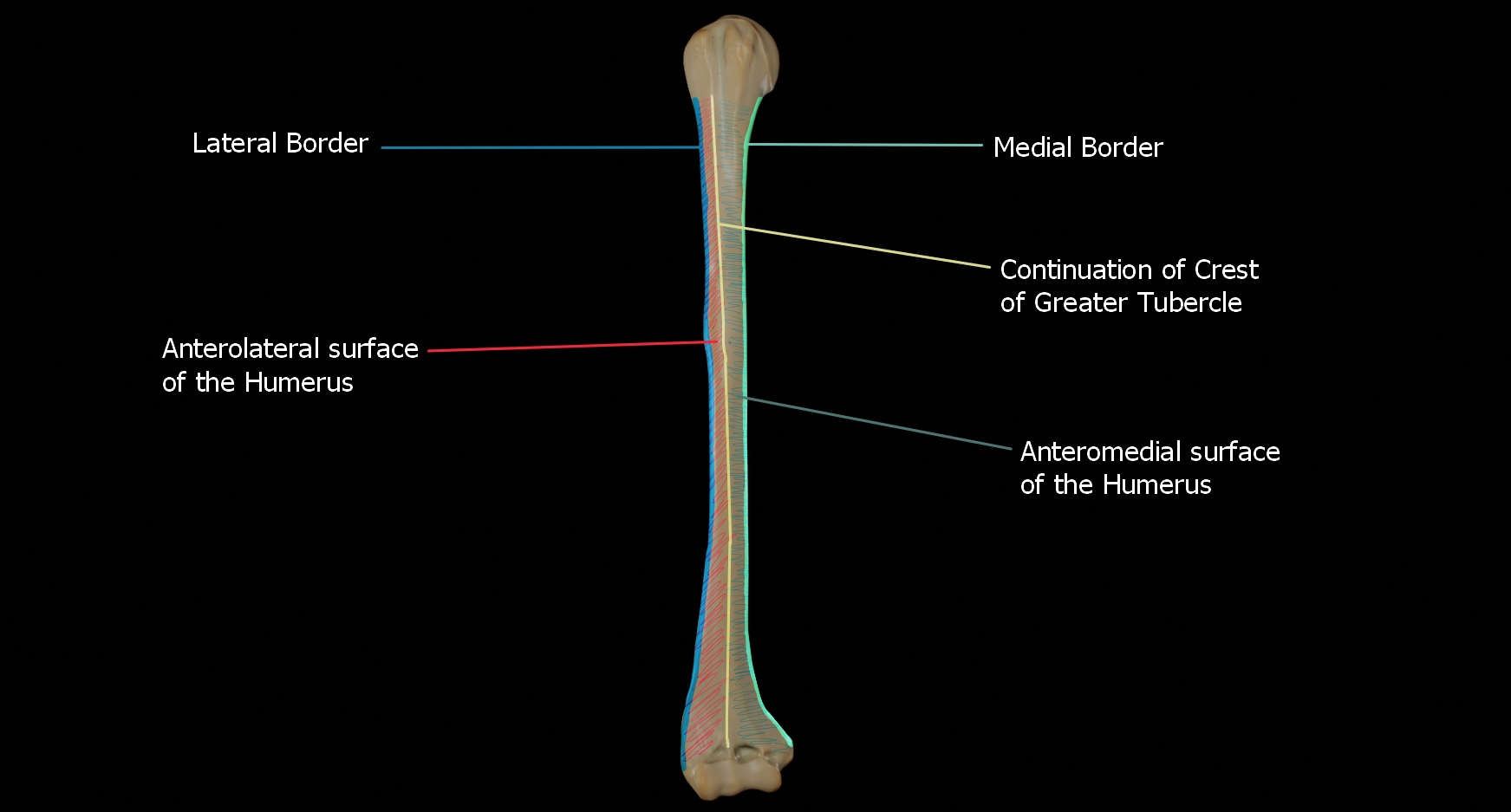
The humerus is the longest and largest bone of the upper extremity, serving as the critical structural foundation for the arm. In clinical 2026 practice, precise identification of labelled humerus landmarks is essential for orthopedic surgeons, radiographers, and physical therapists to ensure accurate diagnosis of fractures, nerve impingements, and soft tissue attachments. This guide provides a detailed breakdown of the humerus, its functional zones, and the clinical implications of its specific anatomy.
Structural Anatomy and Primary Landmarks of the Humerus
The humerus is functionally divided into the proximal end, the shaft (diaphysis), and the distal end. Each region contains specific prominences that serve as attachment points for muscles or transition zones for neurovascular structures. For accurate clinical charting and surgical planning in 2026, healthcare professionals must utilize standardized nomenclature.
The Proximal Humerus
The proximal humerus serves as the anchor point for the shoulder joint (glenohumeral joint). Key anatomical features include:
- Greater Tuberosity: Located laterally, this serves as the insertion point for the supraspinatus, infraspinatus, and teres minor muscles of the rotator cuff.
- Lesser Tuberosity: Located anteriorly, serving as the insertion point for the subscapularis muscle.
- Bicipital Groove (Intertubercular Sulcus): A deep groove separating the two tuberosities, housing the long head of the biceps brachii tendon.
- Surgical Neck: A common site of fracture; it is the constricted area distal to the tuberosities where the humerus narrows.
The Humeral Shaft
The mid-section of the bone is primarily cylindrical but features critical markings for diagnostic orientation:
- Deltoid Tuberosity: A roughened area on the lateral surface of the shaft where the deltoid muscle inserts.
- Radial Groove: A shallow depression running obliquely across the posterior surface, marking the path of the radial nerve. This is a critical zone of caution during internal fixation surgeries, as nerve damage here results in characteristic wrist drop.
Distal Anatomy and Articular Surfaces
The distal humerus transitions into a complex structure forming the elbow joint. Understanding these landmarks is vital for diagnosing common injuries like supracondylar fractures.
- Capitulum: A rounded, lateral knob that articulates with the head of the radius.
- Trochlea: A pulley-shaped medial surface that articulates with the trochlear notch of the ulna.
- Medial Epicondyle: The prominent bony projection on the medial side; it is easily palpated and serves as the attachment point for the forearm flexor muscles.
- Lateral Epicondyle: The smaller projection on the lateral side, serving as the attachment point for the forearm extensor muscles.
- Olecranon Fossa: A deep posterior depression that accommodates the olecranon process of the ulna during full extension of the elbow.
Comparative Overview of Humeral Landmark Significance
The following table summarizes the clinical significance of specific labelled landmarks found during radiographic assessment in 2026 clinical standards.
| Anatomical Landmark | Primary Clinical Association | 2026 Diagnostic Focus |
|---|---|---|
| Surgical Neck | Axillary nerve injury risk | Proximal fracture displacement analysis |
| Radial Groove | Radial nerve palsy risk | Mid-shaft fracture management |
| Medial Epicondyle | Ulnar nerve entrapment | Pediatric supracondylar trauma |
| Capitulum | Radiocapitellar joint stability | Osteochondritis dissecans screening |
| Bicipital Groove | Biceps tendinopathy | Ultrasound-guided injection site |
Clinical Implications for 2026 Orthopedic Care
Modern orthopedic protocols in 2026 emphasize a functional approach to humeral trauma. Treatment strategies have shifted toward early mobilization and, where necessary, minimally invasive osteosynthesis.
Fracture Classification Systems
Clinicians currently utilize the Neer Classification system for proximal humeral fractures, which evaluates the displacement of four segments: the articular surface, the lesser tuberosity, the greater tuberosity, and the humeral shaft. For shaft fractures, the AO/OTA classification remains the gold standard, focusing on the morphology of the fracture line (transverse, oblique, or comminuted).
Soft Tissue and Nerve Considerations
A critical component of the 2026 standard of care is the mandatory documentation of neurological status distal to a humeral injury. Because the radial nerve lies in direct contact with the bone at the radial groove, any humeral shaft fracture requires an immediate check for thumb extension and sensory changes in the dorsal web space of the hand. Failure to document this pre-intervention often leads to preventable malpractice exposure during the post-operative recovery phase.
Troubleshooting and Practical Diagnostic Tips
When analyzing a labelled humerus on an X-ray or CT scan, practitioners should follow these diagnostic steps to ensure no oversight occurs:
- Check the cortical integrity: Look for subtle cortical breaks in the supracondylar region, which can be easily missed in standard anteroposterior (AP) views.
- Evaluate the "Fat Pad" signs: In elbow imaging, displacement of the posterior fat pad is a highly sensitive indicator of an occult fracture, even if the humerus appears intact.
- Assess the alignment of the capitulum: On lateral projections, the capitulum should align with the shaft of the humerus. Any deviation suggests an articular fracture or dislocation.
- Verify hardware placement: If reviewing a post-surgical humerus, ensure that screws are not breaching the articular surface of the trochlea or capitulum, as this will lead to immediate joint degradation.
Frequently Asked Questions
What is the most common site for a humeral fracture? The surgical neck and the mid-shaft (diaphysis) are the most frequent sites of humeral fractures in both adult and pediatric populations. These areas are vulnerable due to the transition between the dense cortical bone of the shaft and the more cancellous bone of the proximal end.
How do you distinguish between the greater and lesser tuberosity? On an AP view of the shoulder, the greater tuberosity is the most lateral point of the proximal humerus, while the lesser tuberosity is located medially, just beneath the humeral head. Their identification is crucial for assessing rotator cuff integrity.
Why is the radial groove clinically important? The radial groove is the anatomical pathway for the radial nerve; fractures involving the mid-shaft of the humerus carry a high risk of iatrogenic or primary nerve injury, leading to wrist drop.
What is the significance of the olecranon fossa in imaging? The olecranon fossa is a key landmark for determining proper elbow extension during imaging; if this area is obscured by edema or hemorrhage, it often indicates an intra-articular fracture.
Is surgery always required for a humeral shaft fracture? In 2026, most closed humeral shaft fractures are managed non-operatively with a functional brace, provided that alignment remains within acceptable parameters (usually less than 20 degrees of angulation).
Expert Consultation and Referral Pathways
If you are experiencing persistent pain or limited range of motion following a humeral injury, it is imperative to consult a board-certified orthopedic surgeon. Ensure your imaging is performed at a facility that utilizes 2026-standard high-resolution digital radiography. If you are covered by an HMO plan, verify that your orthopedic specialist is within your specific network and that you have obtained the necessary primary care referral to prevent out-of-pocket financial liability.